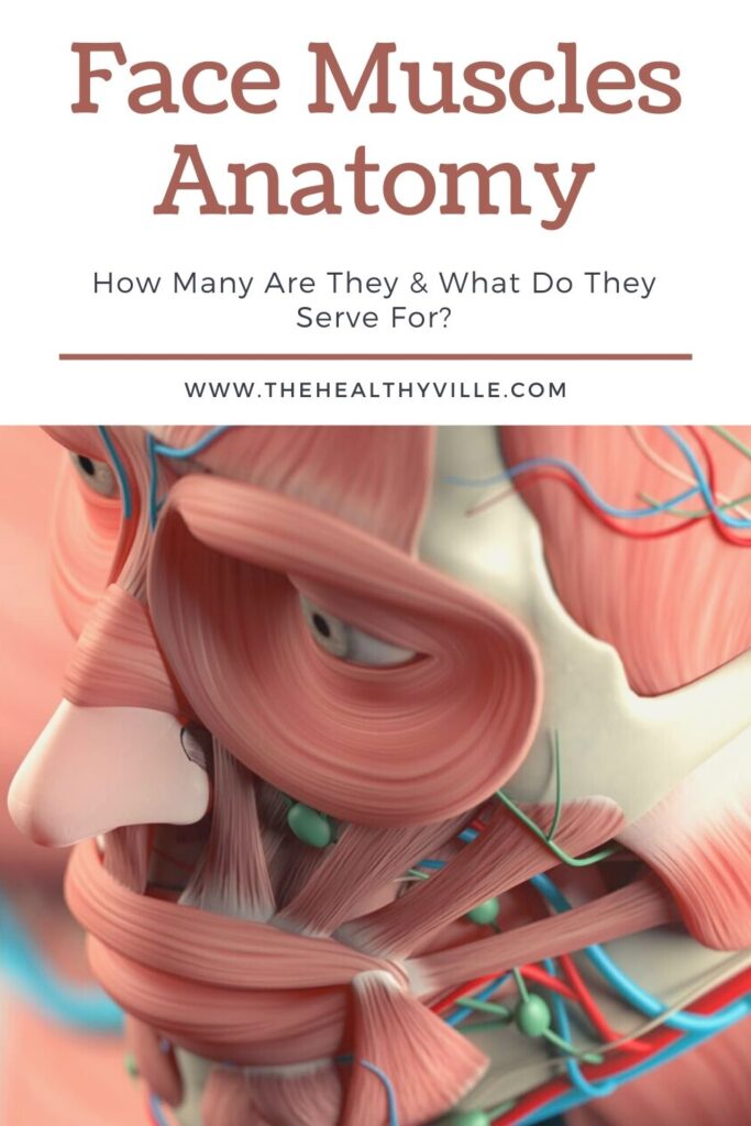
The face muscles anatomy is a pretty incredible one, as there’s a muscle responsible for each part of the face. Make sure you read all about this interesting theme!
We put the muscles of the face into operation when we speak, laugh and eat. Keep reading and know everything that is under the skin of the face, to discover the true face muscles anatomy.
What are the muscles of the face?
Although we do not take them into account, the muscles of the face are very important for expression, that is, for gestural language. As well as for food and relief and the shape of the face.
The muscles of the face are distributed around the eyes, the mouth, the nose or the forehead, even the ears! Even when the latter do not move at will.
Face muscles anatomy
The muscles of the face are between the skin and the bones of the face or skull. Most are flat beams. They are not surrounded by a fascia, with the exception of the buccinator. The fascia is a membrane that allows free movement without friction.
In total, there are more than 40 muscles in the face. They are found around the facial openings (mouth, eyes, nose, and ears). There are also along the forehead, at the temple, at the jaw and at the base of the neck.
We also know them as craniofacial muscles, innervated by the facial nerve, which is the seventh of the cranial nerves. The facial artery vascularizes them.
Functions of the muscles of the face
The muscles of the face fulfill specific and differentiated functions, although they can work in a coordinated way to produce expressions. It can be said that they perform four types of basic tasks:
- They are involved in eating: both in chewing and in tasting food.
- Phonation or speech: moving or modulating the lips, even the nose, since there are nasal sounds, in which part of the air comes out through the nostrils.
- The facial expressions: strangeness, doubt, joy and others involve the movement of various muscles of the face.
- They shape the face: of course, this is influenced by the bones and the texture of the skin.
Mouth muscles
This is the largest group in terms of the muscles of the face. They allow us to open our mouths, move it to one side or the other, project our lips to speak, sing, laugh, eat and even kiss.
Most of them are connected to each other by a fibromuscular axis called a modiolus, into which they insert their fibers. This axis is located at the corners of the mouth, near the corners of the lips.
Buccinator
Its name derives from the Latin buccina (horn). It is the one that brings the corner back, increasing the opening of the mouth.
It also helps us, when whistling or playing a wind instrument, to bulge the cheeks to blow.
Levator anguli oris
This is a thin muscle that is shaped like a sheet, above the upper jaw. As the name implies, its main function is to raise the angles of the lips.
Anguli oris depressant
Contrary to the previous one, it contributes to the expression of feelings of sadness or annoyance, as well as to the opening of the mouth when speaking or eating. It is triangular in shape and is located on each side of the chin.
Major and minor zygomatic
Both muscles run diagonally, from the cheekbone to the corner of the lips. They help raise the angle of the mouth, contributing a variety of expressions.
Square muscle of the chin
Also called lower lip depressor (or depressor labii inferioris). It is, as its name implies, quadrangular in shape and is found on the chin. Roll down the lip, as well as the corners.
Mentalis
It is below the previous one, in the chin area. The mentalis is conical. It helps shape the lips by ingesting fluids, as well as by conveying certain feelings, such as sadness, mockery, or contempt.
Masseter
It is the most important when it comes to eating. This is the king of chewing. It has a rectangular shape and is made up of two fibers, and it goes from the cheekbone to the jaw. It is one of the strongest in the body.
Temporalis
Although seemingly far from the mouth, this fan-shaped muscle on the side of the head (from the temples to behind the ears) helps close the mouth and move the jaw to chew food.
Risorio
It is triangular in shape, found on the sides of the face, with the vertex pointing towards the corners. It performs the opposite function of the buccinator, as it helps to hollow out the cheeks.
Despite its small size, it is very important in terms of the function of vocalizing, helping to produce speech sounds, along with the zygomatic (major and minor) and orbicularis. Also, as its name implies, it is largely responsible for laughter.
Elevator of the upper lip
Its full name is levator labii superioris alaeque nasi. It is a thin, strap-shaped muscle located on both sides of the nose.
It allows to elevate the lip, exposing the upper teeth, deepening or increasing the curvature of the nasolabial or nasolabial fold. Although it is used to smile, it also helps to express contempt.
Orbicular of the mouth
The orbicularis surrounds the entire orbit of the mouth and lips. You have to say their full name so as not to confuse it with the one with the eyes. It is also known as orbicularis oris.
It consists of two parts: a peripheral and a marginal. Both originate from the modiolo. Its function is to produce the movements of the lips, opening and closing, puckering or twisting. Therefore, participate in communication.
Muscles around the eyes
Around the eyes there are almost as many muscles and functions as complex as around the mouth. Let’s see.
Orbicular of the eyes
This muscle of the face is made up of an eyelid and an orbital part. The palpebral area forms the eyelids and the orbital region encloses it concentrically.
The orbicularis oculi is what makes us blink or close our eyes when winking or sleeping. According to experts, the eyelids are kept open because the frontal is stronger than the orbicularis.
On the other hand, the deep palpebral part, which is located towards the lacrimal sac, pulls on the eyelids and lacrimal papillae, dilating the mentioned sac. In other words, this is the muscle that makes us cry.
Superciliary Corrugator
This is a thin muscle. It is located in the internal part or rather under the brow bone. When contracted, it pulls the eyebrows to the middle, contributing to the frown expression.
Other muscles in the eyelid area
In this small area of the face there are other muscles, whose function is related to the movements of the eye, the eyelids and expressiveness. Among these are the following:
- Corrugator: move the eyebrows, raising them or relaxing.
- Elevator of the upper eyelid: works in conjunction with the orbicularis oculi to open the eyes.
Muscles of the nose
There are more muscles in and around the nose than we might believe. Although they are small and with little relative mobility. Most participate in respiratory function and expressiveness.
One of the main ones is the nasal or nasalis. It is located on each side of the back of the nose. It consists of two parts: a wing and a transversal.
Compresses the nasal opening, assisting in deep breathing. To this same extent, it can convey the idea of anger.
For its part, the procerus is a small muscle with a pyramidal or fan shape. It is located on top of the bridge; that is, between the eyebrows. Hence, it is the frown muscle
Other muscles of the nose are as follows:
- Mirtiform – Helps close the nostrils (although only to a certain extent).
- Transverse and dilator: they work together to open or dilate the nostrils.
Ear muscles
In the ears, or rather around, there are muscles too. For some animals they are useful, as it helps them orient the ear towards the sound.
There are exactly 3:
- Auricularis anterior: it is shaped like a triangle, with the apex towards the ear. As its name implies, it is located on the front face of the pavilion.
- Posterior auricularis: it is the smallest. It is made up of two or three fibers that are inserted into the temporal mastoid.
- Auricularis superior: it is in the part of superior of the pavilion, towards the temporal.
Other muscles of the face
In the face there are other muscles, although they are not so visible. And although we do not perceive them when we look in the mirror, they have important functions. Such is the case of the external pterygoid.
This muscle is located in the upper part of the lower jaw. Participates in the chewing process and is responsible for opening the mouth. So it also comes in when we talk, laugh, or yell.
And finally, the occipitofrontal. It is a large muscle, extending from the eyebrows to the occipital bone. The occipitofrontal is the one that produces the wrinkle of the forehead, which is marked when we are surprised.
Exercises the muscles of the face
Our life depends on the proper functioning of face muscles anatomy. That’s how it is. They are very important. We need them to eat, to talk and ask for help, to interact with others.
Although most do not or do not do it very often, it is possible to exercise the muscles as if we were going to a gym. There are, for example, facial yoga routines, which not only tone them, but also help you look young.
And it is that along with dryness, one of the factors that make the face look flabby is the loss of tone in the facial muscles. So smile: smiling is the best exercise for the muscles of the face.
Don’t forget to SHARE the face muscles anatomy with your friends and family on your social networks!

