Subungual hematoma treatment includes lowering the pain and discomfort. Learn about its diagnosis and what to do about it.
Subungual hematoma is the most common cause of brown or black nail pigmentation. The damage is due to a direct hit or crush injury to the distal part of the fingers, for example, by pinching a limb in a doorway.
When blood enters the space under the nail, it puts painful pressure on the nail bed. This is annoying and makes it difficult to perform everyday activities with your hands. The injury may also be accompanied by phalangeal fractures, nail detachment, or fingertip amputations.
What is a subungual hematoma?
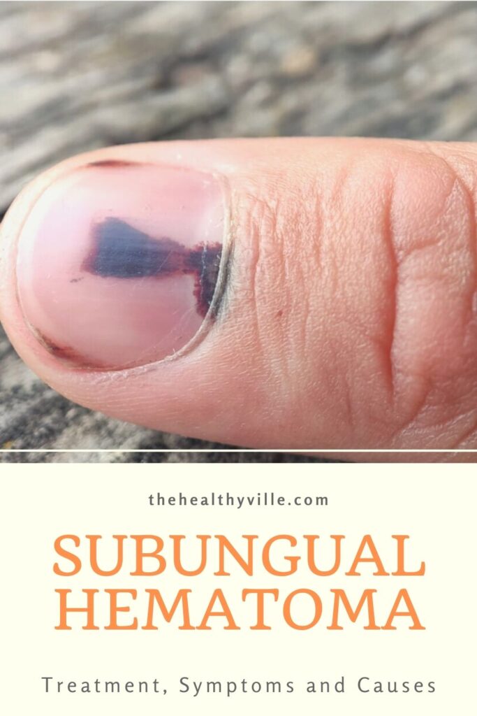
Subungual hematomas are injuries to the nail bed in which bleeding develops under the nail. The usual process is that it begins with a bruise under the tissue, which appears discolored, with round margins on the proximal edge and with a striped distal end with filaments. Although there are variants, this is the most frequent presentation
There may also be one or more small round red and black spots around the main hematoma. These represent smaller outflows of blood from the tiny arterioles that surround the primordial lesion.
What are the symptoms?
Subungual hemorrhage presents as a single pigmented nail that can be painless, tender, or painful. Patients usually attend the medical consultation with discomfort and with a discoloration that is observable with the naked eye.
Soon more signs are associated. Reactive inflammatory changes, such as edema and erythema around the nail fold, may be seen shortly after injury. Edema is the accumulation of fluid and erythema is a slight reddish color.
The color variation of the subungual hematoma is related to the duration and stage of healing. According to studies by Skin appendage disorders, they appear with the following shades:
- Reddish.
- Violet.
- Brown.
- Black.
- A combination of the above.
How is the diagnosis?
The diagnosis of subungual hematoma is, in basic terms, a clinical act of observation. The doctor examines the entire structure of the nail for alterations in the fold and the entire finger to determine motor function, sensitivity and circulation in the area, which could be affected.
Dermoscopic features may include the following:
- Homogeneous or variable colors: reddish, purple, brown or black.
- Rounded shape: with fading on the periphery of the hematoma.
- Periungual hemorrhage: these are small associated bleeds around the nail.
- Linear white marks: on the nail plate, mainly due to the loss of transparency caused by the accumulated blood.
- Yellow staining: This can be registered in the distal region of the nail plate.
Sometimes it is necessary to request an x-ray to evaluate an underlying fracture. Remember that trauma are common causes and a broken phalangeal bone is not an unusual situation.
Differential diagnostics
Subungual hematoma must be differentiated from melanotic pigmentation, that is, from melanoma of the nail. This is not always easy with the naked eye, as bruises are not usually round in shape, but tend to be longitudinal.
The most important role of nail dermoscopy is to allow the recognition of subungual hematoma and differentiate it from the following disorders:
Nail melanoma: Dermoscopic features suggestive of nail melanoma include a brown to black background with longitudinal lines irregular in thickness and spacing.
Fungi: one of the causes of black nail coloring is fungal melanonychia caused by Trichophyton rubrum or other non-dermatophyte fungi. Cases of nail mycosis usually have dull black pigmentation, wider at the distal edge than at the proximal edge, called the reverse black triangle.
Subungual hematoma treatment
A treatment for subungual hemorrhage, in most cases, is not necessary. The key is to make sure the bruise is no more than 48 hours old. In the case of repetitive injury, avoid precipitating factors such as tight or poorly-fitted shoes.
Current recommendations for drainage of acute subungual hematomas (less than 48 hours) advocate simple trepanation. In this procedure, doctors make a hole in the nail to drain and decompress the hematoma.
Recommendations are for the patients to go on a follow-up to ensure that there is no infection and the hematoma is over. Several studies carried out by JAAPA have shown that trepanation has a good cosmetic result and the complication rates are similar in most cases, without being high.
Always consult with the specialist
Subungual hemorrhage resolves slowly over months or even years. Toenails take longer to recover than fingernails and that often scares patients, who assume it should be fixed sooner.
Recommendations are, from a medical point of view, not to try home treatments to avoid superinfection of the area or subsequent complications. In fact, in the event of any discoloration, consult a dermatologist, who will carry out the corresponding evaluation.
Don’t forget to SHARE the subungual hematoma treatment, causes and symptoms with your friends and family on your social networks!